
This is a placeholder text
Group text


by Prager on 20 June 2013 - 14:06
I am in constant study of hips and elbows. I have found this evaluation written on veterinary website. It is a very informative article. But then I have seen this. I would like to run it by you who know about hips a little. What do you think?:
True or False?
Here is the link:
http://www.infovets.com/healthydog/f610.htm
Prager Hans
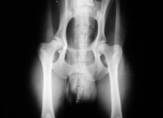
| Figure 16: This is a completely normal pelvis. The hip sockets and head of each femur are also normal. Notice the smooth, rounded head of each femur and how they fit nicely into each hip socket (acetabulum). |
Here is the link:
http://www.infovets.com/healthydog/f610.htm
Prager Hans
by Ibrahim on 20 June 2013 - 14:06
I do not know if it is true or false but I want you to know I will be following this post closely in order to learn, I can say with confidence that so far no matter what I tried I hadn't managed to comprehend anything at all from x-rays. Thanks for starting one more educating post Prager.
Ibrahim
Ibrahim
by Ibrahim on 20 June 2013 - 15:06
I will share what I see but don't turn a laughing cheek on me, from my position and looking at the hips, the left hip in the picture (which is in reality the right hip of the dog) something is not right about the head itself, it looks as if it is worn out (part of its volume was lost), like when you grind a bone and the outer crest gets washed away.
Ibrahim
Ibrahim

by Sunsilver on 20 June 2013 - 15:06
Both hips are partially out of the sockets. The right is worse than the left, but that could be mostly due to the poor positioning.
No sign of degenerative joint disease...YET.
No sign of degenerative joint disease...YET.
by Til on 20 June 2013 - 15:06
The positioning isn´t right, so it seems as if the right hip isn´t correct. In fact both hips are ok.
by Sunsilver on 20 June 2013 - 15:06
My bad...I downloaded the x-ray to my picture file, so I could enlarge it...and I now realize I was looking at the WRONG PICTURE! 
The right hip doesn't look as good as the left, likely due to the poor positioning. I think both hips will get an OFA good rating.
The right hip doesn't look as good as the left, likely due to the poor positioning. I think both hips will get an OFA good rating.
by Blitzen on 21 June 2013 - 08:06
How old? What breed? Sedated? Positioning is not diagnostic.

by Ryanhaus on 21 June 2013 - 09:06

by ziegenfarm on 21 June 2013 - 15:06
jmho, but the positioning in this xray is lousy. as a vet, he should have posted something more flattering to his skills.
i wouldn't want people to see this one. furthermore, this is not a film of a "completely normal" dog. the dog shows
the beginnings of remodeling & calcification----especially in the dog's rt hip. :(
pjp
i wouldn't want people to see this one. furthermore, this is not a film of a "completely normal" dog. the dog shows
the beginnings of remodeling & calcification----especially in the dog's rt hip. :(
pjp
by joanro on 21 June 2013 - 16:06
I agree with Mirasmom and ziegenfarm.
I think TV because the pelvis is asymmetrical and the right side of the pelvis (left side of picture) appears flat, as in TV.
The right hip looks like the neck is thicker than the left and looks like bone spurs on outside of head of femur and neck. The dog's left hip looks ok from here.
If the dog has TV, then better positioning probably can't be attained. JMO.
Contact information Disclaimer Privacy Statement Copyright Information Terms of Service Cookie policy ↑ Back to top