

This is a placeholder text
Group text


by raulgrmn on 28 December 2013 - 04:12
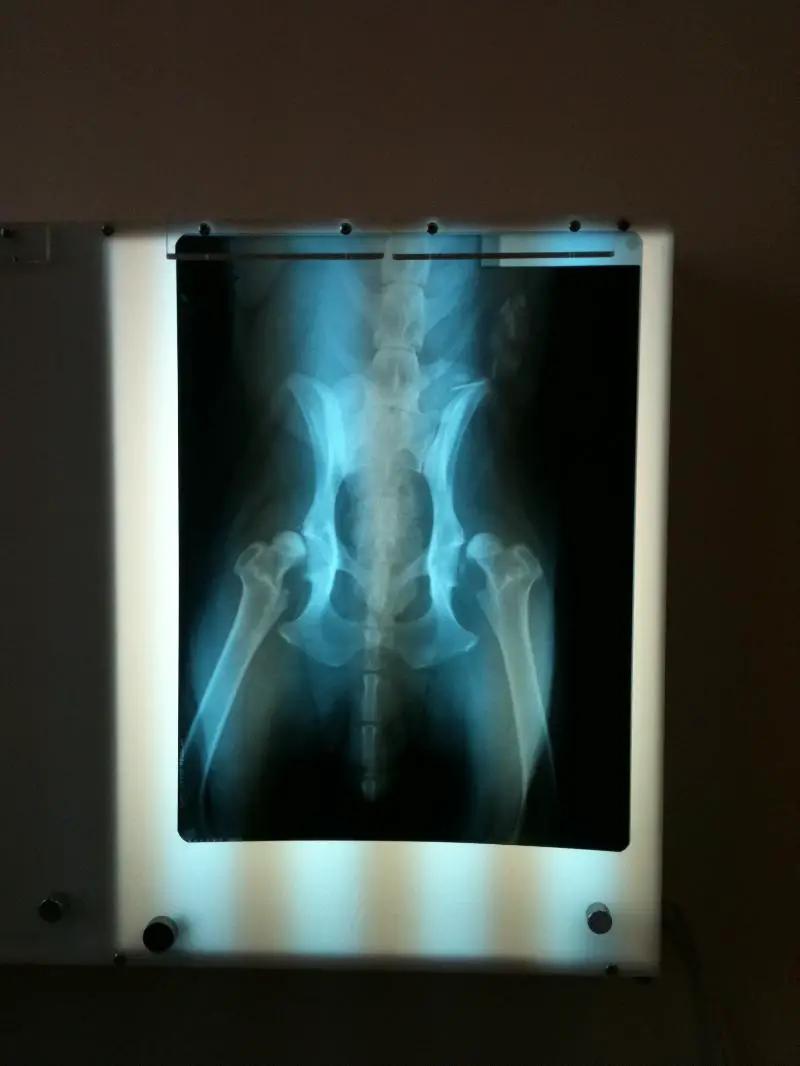
What are your thoughts on these hips (including positioning) 27 month old female GSD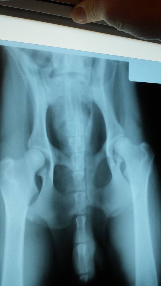
by hexe on 28 December 2013 - 04:12
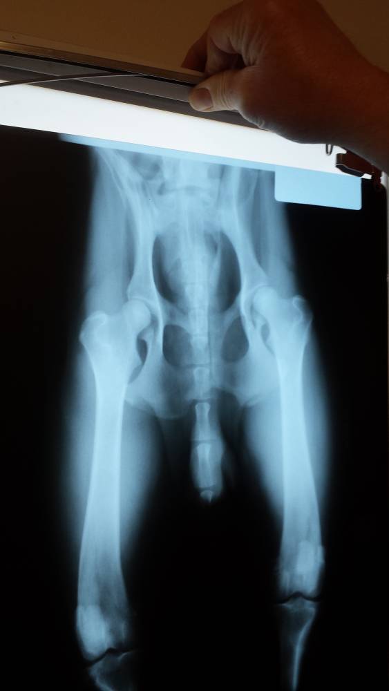
Positioning in second film better than first--don't see anything alarming in either, but the second shot shows how deeply the femoral heads are seated much better. Should go OFA Good.
by Nans gsd on 28 December 2013 - 11:12
I am thinking the right side of the screen (probably left hip) may be a bit shallow, however, hope you get a good but could be fair. Best of luck Nan
PS: can you post results when you receive?? Thank you...
PS: can you post results when you receive?? Thank you...
by raulgrmn on 28 December 2013 - 21:12
Had them done earlier this year and they came back Fair. I was wondering if it would be worth re-taking and re-submitting them. I've seen hips no were good as these get fair and even hips slightly worse/similar get a good rating.
by SitasMom on 28 December 2013 - 21:12
good to fair, don't worry.

by CMills on 28 December 2013 - 22:12
They will pass, I'm guessing at a Fair due to the Left hip not as good as the right. A pass is a pass!!
by raulgrmn on 29 December 2013 - 01:12
Thank you everyone for your opinions. Happy Holidays!

by lawhyno on 03 January 2014 - 21:01
Good read

by GSDNewbie on 04 January 2014 - 00:01
the only thing I see they could have graded against is the shape of the femoral head in the leg left hip. It is also slightly larger and shallower to me. Other than that I have seen lesser hips go good.... I expected a good for these.

by Sunsilver on 04 January 2014 - 00:01
Excellent hips
.jpg)
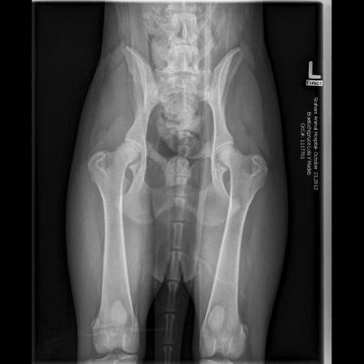
You can start by looking at these two images to learn what good hips look like, but first, you need to learn the different parts of the hip joint. The leg bone (femur) ends in a ball and socket joint, which articulates with the pelvis (the socket part). The part of the femur between the ball of the hip and the actual leg bone is called the neck of the femur. It should be well-defined (narrower than the ball of the hip.) If there is little to no neck on the femur, that means the ball joint is not well developed, and likely does not fit well in the socket.
Here's a set of hips with a poorly developed neck on the femur: The ball of the hip is almost the same diameter as the neck!
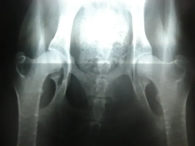
Another thing to look at is how well the ball of the hip fits into the socket. The tighter the fit, the better. Here's a close-up of a nice, tight hip socket:
.jpg)
Notice there is a line that cuts diagonally across the ball of the hip. This marks the edge of the area that actually articulates with the socket of the pelvis. You want as much of this area to be inside the socket as possible. In really bad hips, the ball can be totally dislocated from the socket. If there is a lot of space between the ball and the socket, this is referred to as 'subluxation' of the joint. Total luxation of the joint means all of the ball is outside the socket.
The ball of the hip is not perfectly round. There is a small dimple in it where a blood vessel enters the bone. This is sometimes mistaken for a deformity.
Subluxation of the hips:
.jpg)
Total dislocation:
Next, look at that all-important area that articulates with the pelvis. If it is rough and pitted, or if there are extra spurs of bone, this is going to cause pain. These imperfections in the surface of the joint are referred to as remodellng. This does't show up in very young dogs with bad hips, but it develops with normal everyday wear and tear as the dog gets older.
Other areas of the joint can show arthritic changes, too, but these generally don't cause as much pain as damage to the joint surfaces.
Another important detail is knowing what constitutes a good x-ray. OFA will reject x-rays that are not done to their standards. The dog should be sedated, to relax the muscles. A female should not be x-rayed when she is due to come in heat, as this can cause the ligaments to loosen temporarily. The x-ray must show the knees. The legs should be turned in, and parallel to one another. The pelvis should be absolutely level, and the same amount of the pelvic bone should be covered on both sides by the leg bones. X-rays # 3 and 5 on this page are what you should look for. Although the pelvis in #5 is slightly tilted, it's not enough to affect the reading of the x-ray.
Contact information Disclaimer Privacy Statement Copyright Information Terms of Service Cookie policy ↑ Back to top