

This is a placeholder text
Group text


by Living Fence on 30 October 2015 - 14:10
A video that explains the terminology and what it means at the example of an actual x-ray. I found it helpful. Not sure whether I'd expect an OFA 'good' on it though, I'd expect a 'fair' based on the second hip (slightly incongruent?). But doesn't matter, the point is that it shows what the terminology means with respect to an actual x-ray.
(Don't know why a video like this needs music but there is always a mute button.)
http://www.screencast.com/t/PLJRdOsMudHw
A drawing that explains the OFA ratings:
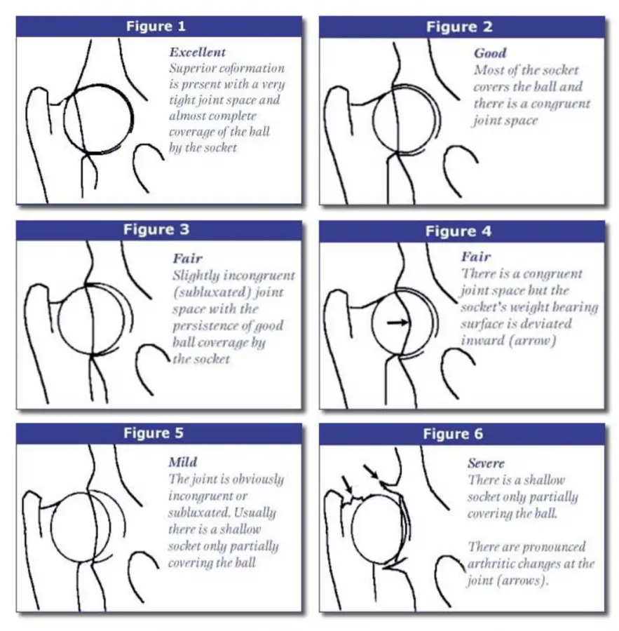
by vonrivera on 11 December 2015 - 02:12
Vonrivera

by srfwheat on 11 December 2015 - 03:12

by SimbaE on 11 December 2015 - 07:12
by Nans gsd on 14 December 2015 - 22:12
Thx for sharing the video though, very informative. Nan

by Hundmutter on 18 December 2015 - 08:12
should see this.
And yes please, as already asked ^^^, can anyone put up
any similar diagrammatic explanation of ED ?
We may disagree as to the best method of checking the
joints; we may argue about the causative factors involved
in joints not being healthy - but we can all at least take in
the information in Figures 1 thru' 6 above, as to what constitutes
a well-formed joint as distinct from a poor one.
by srfwheat on 19 December 2015 - 04:12
Like Hundmutter, I would like to see a diagrammatic explanation on elbow dysplasia similar to the diagram originally posted by Living Fence on hip dysplasia. The diagram really helps in the understanding of where some of the gradings by OFA are coming from. Thank you again for the helpful diagram.
by Living Fence on 19 December 2015 - 15:12
Unlike HD, ED is not diagnosed based on degrees of incongruence in the joint, and thus there is no diagram for ED that is equivalent to the one above on HD. Instead the ratings for ED refer to degree of degeneracy of the bones and cartilage in the joint, from zero to severe.
For that reason, ED ratings are meaningful only at an age when degenerative change can have taken place and can be seen in an x-ray, two years of age.
The elbow joint involves three bones, and there are four different types of degenerative condition, all affecting different sites in the elbow joint. There are diagrams of canine elbow anatomy, as well as drawings of the different degenerative conditions. I found the OFA's explanation and images very educational, as well as one other page:
by Hundmutter on 19 December 2015 - 16:12
so I can have it to hand; now considerably clearer
about just what ED can amount to & what we're looking
for.
by srfwheat on 20 December 2015 - 07:12
Living Fence - Thank you for the information and links on ED.
Contact information Disclaimer Privacy Statement Copyright Information Terms of Service Cookie policy ↑ Back to top