
This is a placeholder text
Group text


by Ibrahim on 10 December 2012 - 17:12
Ibrahim
From Wikipedia
Hip dysplasia is an abnormal formation of the hip socket that, in its more severe form, can eventually cause crippling lameness and painfularthritis of the joints. It is a genetic (polygenic) trait that is affected by environmental factors. It can be found in many animals and occasionally in humans, but is most commonly associated with dogs, and is common in many dog breeds, particularly the larger breeds.
Hip dysplasia is one of the most studied veterinary conditions in dogs, and the most common single cause of arthritis of the hips.[1]
Normal hip anatomy
In the normal anatomy of the hip joint, the root (the thigh bone) is connected to the pelvis at the hip joint. The almost spherical end of the femurhead (the caput, or caput ossis femoris) fits into the acetabulum (a concave socket located in the pelvis). The bony surface of the femur head and of the acetabulum are covered by cartilage. While bones provide the strength necessary to support body weight, cartilage ensures a smooth fit and a wide range of motion. Normal hip function can be affected by congenital conditions such as dysplasia, discussed in this article, trauma, and by acquired diseases such as osteoarthritis and rheumatoid arthritis.
[edit]Dysplastic hip anatomy
In a hip suffering from dysplasias, two things are commonly abnormal. First, the caput is not deeply and tightly held by the acetabulum. Instead of being a snug fit, it is a loose fit, or a partial fit. Secondly, the caput or acetabulum are not smooth and round, but are misshapen, causing abnormal wear and tear or friction within the joint as it moves.
The body reacts to this in several ways. First, the joint itself is continually repairing itself and laying down new cartilage. However cartilage repair is a relatively slow process, the tissue being avascular.
So the joint may suffer degradation due to the abnormal wear and tear, or may not support the body weight as intended. The joint becomesinflamed and a cycle of cartilage damage, inflammation and pain commences. This is a self-fueling process, in that the more the joint becomes damaged, the less able it is to resist further damage. The inflammation causes further damage. The bones of the joint may also developosteoarthritis, visible on an X-ray as small outcrops of bone, which further degrade the joint.[2]
The underlying deformity of the joint may get worse over time, or may remain static. A dog may have good X-rays and yet be in pain, or may have very poor X-rays and apparently almost no problems. The hip condition is only one factor to determine the extent to which dysplasia is causing pain or affecting the quality of life. In mild to moderate dysplasia it is often the secondary effects of abnormal wear and tear or arthritis, rather than dysplasia itself, which is the direct causes of visible problems.
Causes and effects

In canines, it can be caused by a femur that does not fit correctly into the pelvic socket, or poorly developed muscles in the pelvic area. Large and giant breeds are most susceptible to hip dysplasia (possibly due to the BMI of the individual animal[3]), though, many other breeds can suffer from it. For a list of top 100 breeds affected, by percentage, visit the OFFA Here: http://www.offa.org/stats_hip.html. Cats are also known to have this condition, especially Siamese.[4]
To reduce pain, the animal will typically reduce its movement of that hip. This may be visible as "bunny hopping", where both legs move together, or less dynamic movement (running, jumping), or stiffness. Since the hip cannot move fully, the body compensates by adapting its use of the spine, often causing spinal, stifle (a dog'sknee joint), or soft tissue problems to arise.
The causes of hip dysplasia are considered heritable, but new research conclusively suggests that environment also plays a role.[5] To what degree the causality is genetic and what portion environmental is a topic of current debate. Environmental influences would include overweight condition, injury at a young age, overexertion on hip joint at a young age, ligament tear at a young age, repetitive motion on forming joint (i.e. jogging with puppy under the age of 1 year). As current studies progress, greater information will help provide procedures to effectively reduce the occurrence of this condition.
In dogs, the problem almost always appears by the time the dog is 18 months old. The defect can be anywhere from mild to severely crippling, and can eventually cause severe osteoarthritis.[6]
It is most common in medium-large pure bred dogs, such as Newfoundland Dogs, German Shepherd Dogs, Labrador or Golden retrievers,rottweilers and mastiffs, but also occurs in some smaller breeds such as spaniels and pugs and occasionally (usually with minor symptoms) incats.
[edit]Clinical detection and testing
[edit]Symptoms

"Traditionally, the signs of hip dysplasia are rarely extreme. Usually, only mild to moderate lameness is noted which may suddenly worsen. Dogs with a cranial (anterior) cruciate ligament tear typically hold the affected leg up (which is unusual with hip dysplasia). Patients with back (spinal) problems often scuff their toenails when walking, have an uncoordinated gait, and are weak in the rear limbs. They may be very painful if they have a disc rupture (sciatica) or show no spinal pain in certain degenerative spinal cord conditions (German Shepherd myelopathy)."[7]
Dogs might exhibit signs of stiffness or soreness after rising from rest, reluctance to exercise, bunny-hopping or other abnormal gait (legs move more together when running rather than swinging alternately), lameness, pain, reluctance to stand on rear legs, jump up, or climb stairs,subluxation or dislocation of the hip joint, or wasting away of the muscle mass in the hip area. Radiographs (X-rays) often confirm the presence of hip dysplasia, but radiographic features may not be present until two years of age in some dogs. Moreover, many affected dogs do not show clinical signs, but some dogs manifest the problem before seven months of age, while others do not show it until well into adulthood.
In part this is because the underlying hip problem may be mild or severe, may be worsening or stable, and the body may be more or less able to keep the joint in repair well enough to cope. Also, different animals have different pain tolerances and different weights, and use their bodies differently, so a light dog who only walks, will have a different joint use than a more heavy or very active dog. Some dogs will have a problem early on, others may never have a real problem at all.
Each case must be treated on its own merits, and a range of treatment options exist.
Long term pain
A dysplastic animal has probably lived with the condition since it was only a few months old, and has therefore grown up taking the chronic painfor granted and has learned to live with it. Dogs suffering such pain do not usually exhibit acute signs of pain. Sometimes, they will suddenly and abnormally sit down when walking, or refuse to walk or climb objects which they usually would, but this can equally be a symptom of many other things, including a thorn in the paw, or a temporary muscle pain. So pain recognition is less common a means of detection than the visible gait and other abnormalities described above.[citation needed]
[edit]
[edit]
by Ibrahim on 10 December 2012 - 17:12
Diagnosis
The classic diagnostic technique is with appropriate X-rays and hip scoring tests. These should be done at an appropriate age, and perhaps repeated at adulthood - if done too young they will not show anything. Since the condition is to a large degree inherited, the hip scores of parents should be professionally checked before buying a pup, and the hip scores of dogs should be checked before relying upon them forbreeding. Despite the fact that the condition is inherited, it can occasionally arise even to animals with impeccable hip scored parents.
In diagnosing suspected dysplasia, the x-ray to evaluate the internal state of the joints is usually combined with a study of the animal and how it moves, to confirm whether its quality of life is being affected. Evidence of lameness or abnormal hip or spine use, difficulty or reduced movement when running or navigating steps, are all evidence of a problem. Both aspects have to be taken into account since there can be serious pain with little X-ray evidence.
It is also common to X-ray the spine and legs, as well as the hips, where dysplasia is suspected, since soft tissues can be affected by the extra strain of a dysplastic hip, or there may be other undetected factors such as neurological issues (e.g. nerve damage) involved.
There are several standardized systems for categorising dysplasia, set out by respective reputable bodies (Orthopedic Foundation for Animals/OFA, PennHIP, British Veterinary Association/BVA). Some of these tests require manipulation of the hip joint into standard positions, in order to reveal their condition on an X-ray.
Conditions which can mimic or replicate the symptoms of hip dysplasia
The following conditions can give symptoms very similar to hip dysplasia, and should be ruled out during diagnosis:
- Cauda equina syndrome (i.e. lower back problems)
- Cranial (anterior) cruciate ligament tears
- Other rear limb arthritic conditions[7]
- Osteochondritis dissecans and elbow dysplasia in the forelimbs are difficult to diagnose as the animal may only exhibit an unusual gait, and may be masked by, or misdiagnosed as, hip dysplasia.[8]
It is also worth noting that a dog may misuse its rear legs, or adapt its gait, to compensate for pain in the forelimbs, notably osteoarthritis,osteochondritis (OCD) or shoulder or elbow dysplasia, as well as pain in the hocks and stifles or spinal issues. It is important to rule out other joint and bodily issues before concluding that only hip dysplasia is present. Even if some hip dysplasia is present, it is possible for other conditions to co-exist or be masked by it.
by Ibrahim on 10 December 2012 - 17:12
Assessing the dysplastic hip
Millions of dollars have been expended over the past 50 y in the diagnosis, assessment, and treatment of canine hip dysplasia (CHD). Very little has been resolved in terms of reducing the incidence of the problem, determining how best to diagnose it, or even agreeing on a definition of CHD!
Most would agree that CHD is a genetically determined, multifactorial developmental abnormality of the coxofemoral joint in which the hip does not fit together properly (1). Hip laxity produces abnormal development of the femoral head and acetabulum resulting in degenerative joint disease and varying degrees of pain and dysfunction (1,2).
Diagnosis of the condition relies on history of hind limb pain or dysfunction and physical examination findings compatible with hip laxity (positive Ortolani sign or Barden’s test) or degenerative joint disease. Ultimately, a diagnosis of CHD relies on radiography…and that’s when the fun begins!
Even the most casual observer of canine hip radiographs will be able to pick out a severely subluxated coxofemoral joint or one with advanced degenerative changes; however, the detection of early hip changes can be a much more challenging proposition. Breeding decisions or, in some cases, even life or death choices for specific animals can hinge on relative radiographic subtleties.
Veterinarians and dog owners or breeders have 2 major choices when it comes to formal radiographic assessment of their dog’s hips. Certification is provided by the Orthopedic Foundation for Animals (OFA), or hip laxity assessment can be obtained from the University of Pennsylvania Hip Improvement Program (PennHIP).
The OFA (www.offa.org) was founded in 1966 as a not- for-profit organization primarily through the efforts of the Golden Retriever Club of America and the German Shepherd Dog Club of America. The OFA currently maintains registries of several inherited diseases in dogs and cats, including elbow dysplasia, hypothyroidism, congenital cardiac abnormalities, as well as hip dysplasia (2).
Due to its status as the oldest method of assessing dysplastic hips, and because of its origins and ongoing close relationship with the American Kennel Club and individual breed organizations, the OFA enjoys a position of prominence over its newer rival — PennHIP.
And “rival” is the correct word! Owing to personalities on both sides, the interaction between the OFA and PennHIP hasn’t exactly been collegial!
The OFA provides an assessment of a dog’s coxofemoral phenotype leading to “certification.” While radiographs from dogs under 24 mo of age can be submitted for an opinion, certification will not be provided until radiographs are taken at or after 24 mo. A hip-extended ventrodorsal radiograph of diagnostic quality, marked to identify the animal and veterinarian, and properly positioned including both ilia and stifles is required. The OFA advises that the radiographs be taken with the animal under sedation or general anesthesia but there is no requirement that this be done. Submissions to the OFA are blindly assigned to 3 veterinary radiologists who grade the hips into one of 7 categories: excellent, good, fair, borderline, or mildly, moderately, or severely dysplastic. The former 3 categories are eligible for OFA certification (2).
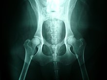
The PennHIP method (www.pennhip.org) was developed by Dr. Gail Smith at the University of Pennsylvania in 1983 with the current radiographic submission system dating back to 1993. PennHIP is an objective measurement of hip laxity, which is considered to be the root of degenerative hip changes in later life. The method has several key differences from the OFA approach. 1) puppies as young as 16 wk of age can be assessed through this method with a high degree of correlation to results obtained at a later age. This is of critical importance to those wishing to breed or begin a dog’s working career before 2 y of age. 2) the PennHIP radiographs must be taken with the animal under anesthesia or heavy sedation to overcome the effects of muscle tension on the assessment of hip laxity. 3) the hips are positioned in a standing angle. The hip-extended position used for OFA radiographs tightens up the joint capsule which has the effect of bringing the hip into closer alignment. 4) a “distracted” radiographic view (Figure 1) is taken using the PennHIP distractor. This device acts as a fulcrum against which the proximal femurs are levered to demonstrate the degree of laxity in the hip (Figure 2). 5) PennHIP radiographs can only be taken and submitted by veterinarians or technicians certified to do so after having taken a training course and submitting trial radiographs demonstrating their proficiency with the technique. 6) all radiographs taken with the PennHIP method must be submitted for inclusion in the database regardless of how abnormal they may appear. 7) the PennHIP analysis does not confer “certification” but rather a “distraction index” (DI), which is an objective measure of the degree of hip laxity, is calculated.

Figure 1
A radiograph of the coxofemoral joints is taken using the PennHip distractor as a fulcrum against which the proximal femurs are levered to demonstrate hip laxity.

Figure 2
The distracted radiographic view demonstrating hip joint laxity. From this view the distraction index (DI) is calculated and compared to other DI’s for the breed.
The DI correlates with the likelihood of the animal developing future degenerative changes in its hips. The PennHIP report also compares the dog to others of the same breed in the database, since DI’s tend to be somewhat variable and breed- specific. PennHIP suggests that breeding stock be taken from dogs that are at least in the top 50% of favorable DI readings for the breed. Given this aspect of the report, the requirement for mandatory submission of films becomes self-evident. It is estimated that perhaps as much as 50% of the OFA films taken are never submitted if there is obvious evidence of dysplasia and little chance of certification (3).
As indicated earlier, these 2 organizations have frequently clashed throughout their histories. The OFA claims that their efforts have resulted in a steady decline in the incidence and severity of CHD and an increase in “good” to “excellent” hips from 1972–2000 (2,4). PennHIP credits the OFA with some impact on the severity of the problem, but points out that due to the lack of mandatory submission of OFA films, any change in the incidence of CHD is unknown (3,5). The OFA radiologists agree that a dog is “normal,” “borderline,” or “dysplastic” almost 95% of the time, and agree on the specific rating almost 75% of the time (2). PennHIP disputes this level of agreement and points out that other “non-OFA” board certified radiologists don’t show anywhere close to this level of agreement in reading hip films. The conclusion they draw is that the OFA method is handicapped by being subjective (3). While the OFA agrees that the PennHIP method is an objective measure, they do not accept the significance of this measurement. In the OFA guide they state, “The degree of joint laxity as demonstrated by… using a fulcrum/stress device — that can be normal, and what degree is abnormal is unknown.” (2) While both sides agree that “tighter” hips seem to be better hips, the OFA points out that not all dogs with demonstrated loose hips go on to develop osteoarthritis (2). PennHIP counters that a Nestlé Purina study demonstrated 55% of dogs certified “normal” by the OFA went on to develop hip osteoarthritis and breedings of OFA normal German shepherds have been able to produce no better than 19% dysplastic pups (3)…and on and on it goes!
What does all this mean for the small animal practitioner and his or her clients? First, it means that the laws of sensitivity and specificity of scientific tests apply very well to OFA and PennHIP radiographs; neither is perfect! In general terms, because of the nature of OFA radiographic positioning and the subjectivity of the test, some false negative results will be produced allowing dysplastic animals into the gene pool. On the other hand, the fact that not every dog with loose hips develops osteoarthritis means that PennHIP assessment will produce some false positives removing some animals unnecessarily from breeding lines. The OFA provides a well-respected, widely recognized standard for diagnosing CHD. PennHip provides a method that can be used much earlier in the animal’s life and implies some conclusions about the dog’s coxofemoral genotype since DIs have been shown to be heritable. There would appear to be room for both methods in the evaluation of dysplastic hips.
I do not understand the article very well especially the diagrams.
by Ibrahim on 10 December 2012 - 17:12
Bony Spurs?
Flat Socket?
Ball irregular
How doe you determine each one of the above, what tells you there is Bony Spurs/Flat socket/Irregular ball when looking at a specific x-ray?
by Ibrahim on 10 December 2012 - 17:12
by Ibrahim on 10 December 2012 - 18:12
I do not understand fully what he explained in the video
And what is Pelvis has to do with hip dysplasia?
by Hundmutter on 10 December 2012 - 18:12
UK British Veterinary Association panel of assessors always say:
breeders & owners should leave it to them to read the hip Xrays sent
to them; too many mistakes made by individuals and/or their
Vets, who think they can read them properly, but can't really. I don't
know if that is true, or if it is the old 'the Doctor is a God' routine ?
I do admit I don't entirely understand the subtle differences why they
allocate the scores to the angles, under the UK's system.
I have seen many Xrays, mostly taken with reasonably good position
-ing, some with sedation and some not. And since visiting PDB a lot
more photos of radiographs. As a rough estimate, I'd say you can
certainly get an idea of hips that are very good or very bad, just by
having seen a number you know were good and comparing others
against those. The video you posted shows that basic assessment
quite well. There is no hiding a 'ball' (femur top) hanging right out of
its 'socket'; nor seeing a pelvis where the bones are clearly so badly
formed that there ARE no sockets to speak of. The difficulties come
with the degrees in between. The giveaway of additional bone growth
at the joints as symptomatic of arthritis is sometimes present, some-
times not. May account for some discrepencies we get between the
different schemes where a dog has been assessed more than once ?
by Ibrahim on 10 December 2012 - 18:12
On another thread some one said something like this " if you're a breeder and you can't evaluate x-rays then you're not eligible to be one"
Ok I am not a breeder but I'd love to at least know whay you guys look at in a x ray, I am lost with all the info on the videos
by Ibrahim on 10 December 2012 - 18:12
by Hundmutter on 10 December 2012 - 19:12
lines on them.
In that last video, where he shows that there were two Xrays of the
same dog, one was badly placed (femurs turned out, kneecaps
turned in, pelvis higher on the table one side), the other was much
better positioned, and got OFA 'Good'. So now you know the 2nd
pic shows you hips graded Good (even if not perfect !), cos the
dog is lying correctly; and that positioning means the femurs go
straight up to the hip joint both sides, and the heads of the bones
are therefore both positioned inside the 'cup' of the joint socket
each side of the pelvis. Where they should normally sit if the dog
was up and walking.
So if you keep that 'view' in your head and then look at another Xray
you should be able to see a) if the positioning is correct; and assuming
it is, you can go on to look at b) do the joints look like a normal ball and
socket arrangement, with the femoral head fitting smoothly into the cup
each side. Or does one or both of them look like it is somewhat out of
position, have irregular shape to the bone (e.g.'flattened' head), or show
big gaps around it.
Now go back to 3/4 way through the Vetstoria podcast video, where he
compares good and bad and gives diagnosis on the beagle. You should
see the differences more now ?
Bony spurs are part of the extra growth that comes with arthritis, and can
be on the rounded head of the femur; or on the spinal vertebrae, so they
might not show in a hip Xray.
Where muscles and ligaments do not hold properly formed joints together
in their correct position, or the bones of the joint are not properly formed,
there is enough movement / wriggle-room for the bones to grind together
(aided by the fact that the synovial fluid in the joint becomes reduced) and
that means that arthritic change to the bone starts to build. Or that bone
gets worn away. And then you may start to see differences on the outside
of the dog (limping; or getting 'stuck' in position, etc...)
Any more precise questions on what you see or don't see ? I'll try.
Contact information Disclaimer Privacy Statement Copyright Information Terms of Service Cookie policy ↑ Back to top