

This is a placeholder text
Group text


by Nans gsd on 08 May 2018 - 19:05
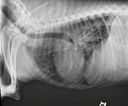
Sorry about the previous posts we could not supersize. OK does this look familiar to anyone; this guy is having a hard time breathing; vets are scratching their heads. Vets as in 4 have looked. I bragged about this forum and said someone on the forum will know. I am thinking cancer in chest. Thank you in advance. Nan

by joanro on 08 May 2018 - 21:05

by Jessejones on 08 May 2018 - 21:05
An ultra sound would give more details on that area.
Add: No, it does not look good, l agree with what Joan and you guessed, but I will keep my fingers crossed for you and Boomer.

by Sunsilver on 08 May 2018 - 22:05
Yes, those white spots look like mets to me. Not normal. Heart looks to be enlarged, too, which would explain the SOB.
Here's a normal chest x-ray of a pregnant bitch:


by emoryg on 09 May 2018 - 00:05
by Nans gsd on 09 May 2018 - 00:05
Joanro he's 12 May 23; in really bad shape and I really don't think he would make it thru surgery?
At Sunsilver: I can't see the comparison except gut, I think. Nan
by Jessejones on 09 May 2018 - 00:05
Undiagnosed Pneumonia may be a possibility as well.
Added:
I am so sorry to hear he is not doing well, Nan. Saw that after I posted.
The ultrasound is not invasive and if he is calm, can be done when he is awake and depending on the vet. He will need his fur shaved though.
The ultrasound can scan his spleen and liver as well for lesions.
Ultrasound is not good for bone or air filled areas, but can show all tissue, including pneumonia signs in the lung.
by joanro on 09 May 2018 - 01:05
by Sunsilver on 09 May 2018 - 06:05
Nan, the roundish white thing you see inside the dog's ribcage is the heart. It's a lot bigger than the one in the second x-ray. Above the heart are a bunch of white dots with dark centers, connected by whitish fibrous strands. That's lung tissue. You normally shouldn't be able to see the lungs on an x-ray, and that whitish tissue should not be there. The lung tissue is also making the heart look blurry compared to the second x-ray. Again, this is because the lung tissue is abnormal, and is partially blocking the x-rays.
by Nans gsd on 09 May 2018 - 14:05
Thank you sunsilver for spelling it out about how to read the x-ray, as this is more explanation than the vets gave us
However Sunsilver I WAS able to see puppy spines in your x-ray.
This is a very special service dog to boot.
Thank you all again and yes we will of course keep him as comfortable as possible. Nan
Contact information Disclaimer Privacy Statement Copyright Information Terms of Service Cookie policy ↑ Back to top